BASIC EYE ANATOMY: All the Parts of the Human Eye at a Glance

In this blog I list a few parts of your visual system, as well as the basic anatomy and physiology.
This information will help you to understand diseases that affect your eyes, as well as the tests we perform to help detect conditions of the eyes.
You’ll find details on select diseases in future blogs. Check ‘em out – if you dare!
To get things started, check out this basic eye anatomy video:
Eyelids
Eyelids are the loose floppy pieces of tissue in front of your eyes. Eyelids are made up of muscle, glands, and connective tissue. Are they important? Absolutely! We have them for four very important reasons:
Protection: The eyelids act as a shield against insects, fingernails, and an assortment of other objects that have the sole purpose of hurting your eyes. The eyelids do a wonderful job preventing potential disasters.
Tear drainage: Each time that you blink (or wink), tears get pooled where the upper and lower lids meet together medially (on the side where your nose is). All the garbage in your tear film – dirt, dust, makeup, old cells, parts of insects, stuff that causes allergies – gets drained away down your nasolacrimal drainage system. Want a real-world analogy? Go flush the toilet.
Tear distribution: You wouldn’t spend any time outdoors without putting sunscreen on. Right? You put a glob of sun stuff in your palm, and then spread it evenly over your body (oooh baby). The eyelids do the same thing with the tear film each and every time you blink. Tears are important to maintain proper eye health which promotes clear vision.
Tear production: The eyelids contain special glands and cells that produce tears. Without them, our eyes would be dry and irritated, vision would suck, and you wouldn’t be able to wear contact lenses. Bummer.
Extraocular muscles (EOM)
If you can look up, down, right, left, and all around, then you can thank your EOMs. Here’s what you need to know:
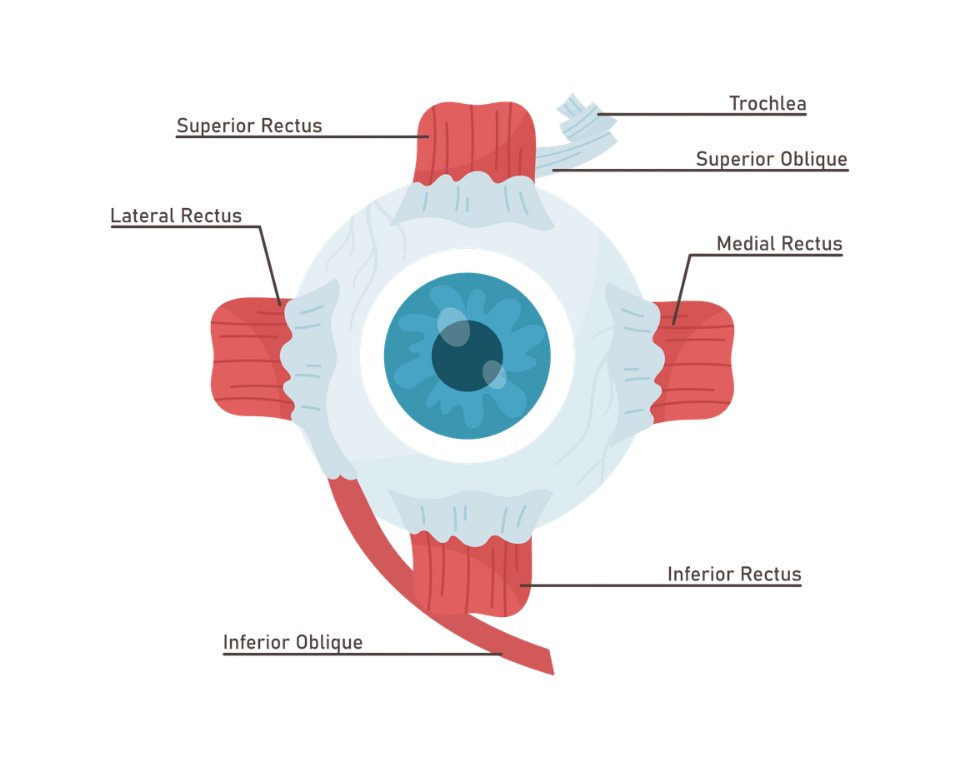
There are six EOMs for each eye. Allow me to introduce you..
Superior rectus: You see that bird up in her nest? Good. Your superior rectus is responsible for elevation of your eye. Otherwise known as “looking up”.
Inferior rectus: Darn. I just dropped that tiny screw that belongs in the hinge of my glasses. Now where did it go? Yup, your inferior rectus is responsible for depression (sigh…) which means that you’re looking down. Now where did that screw go?
Medial rectus: When you look directly to the left, the medial rectus makes your right eye turn in the direction of your nose. This is called adduction. (The same goes for your left eye when you look to your right.)
Lateral rectus: When you look directly to the left, the lateral rectus makes your left eye turn away from your nose. This is called abduction. (The same goes for your right eye when you look to the right).
Superior oblique: Makes the right eye look down when you look left, and makes the left eye look down when you look to the right.
Inferior oblique: Makes the right eye look up when you look to the left, and makes the left eye look up when you look to the right.
Some extra info about your EOM’s:
The oblique muscles also rotate slightly when the eyes abduct (look away from the nose).
The EOMs can move at top speed. They are the fastest muscles in your body. When you look at the world around you, your eyes are moving in small quick movements called saccades. This movement can take place in as little as 1/5th of a second.
The EOMs are 100x stronger than they need to be to move an organ as light in weight as the human eye. This allows for accurate, lightning-fast movement.
EOMs are resistant to fatigue. They have a high nerve-to-muscle ratio compared to other skeletal muscles in the body.
Even though they are highly susceptible to Grave’s disease, where a hormonal imbalance causes swelling and restricted movement of the EOMs, they are often spared in other types of skeletal muscle disease.
The EOMs move about 100,000x per day. This constant movement requires lots of energy, so eat your fruits and veggies and get plenty of sleep.
Conjunctiva
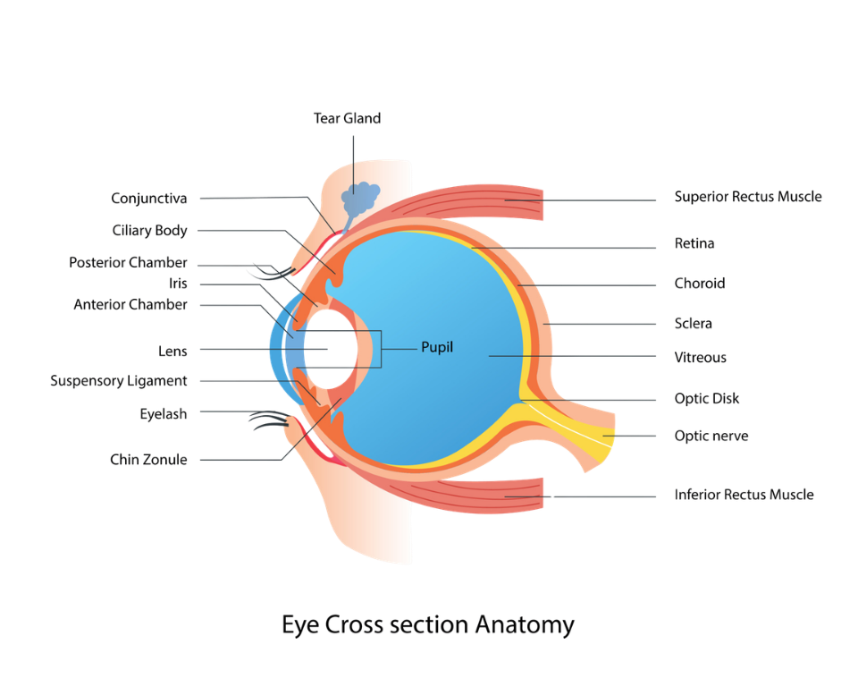
Take a look in the mirror. You see the whites of your eyes? The white structure of the eye is called the sclera. The sclera (and the inside lining of your eyelids) is covered by a thin, translucent membrane called the conjunctiva.

Look in the mirror again. Are your eyes red? If they are, then I hope you had a good time last night. There are plenty of blood vessels embedded in the conjunctiva. When our eyes are dry, irritated, or itchy, the blood vessels dilate. Hence, red eyes.
The conjunctiva also has glands and cells that produce tears, just like the eyelids. When our eyes become infected with too much bacteria, the conjunctiva becomes inflamed.
In the clinic, we call this conjunctivitis.
You call it pink eye.
Everyone calls it disgusting.
Cornea
If you get poked in the eye and it hurts a little, then you probably have an irritated conjunctiva. If you get poked in the eye and you scream HOLY SHIT THAT HURTS! And the pain doesn’t go away, then congratulations, you just found your cornea.
Your cornea is the clear window in the front of your eye. You can see the image of your colored iris through the cornea. You can put a contact lens on your cornea. And when the cornea becomes poked, prodded, irritated, or infected, then it hurts...a lot.
The cornea is made up of several layers of specialized cells and tissue. These layers serve to transmit and bend light into your eye. The cornea is the principal refracting component of your eye, thereby helping the lens inside of your eye to focus light onto your retina.
The nerves of the cornea are very sensitive. If any object gets close to the eye, the eyelids automatically shut in order to avoid any potential damage. Those same nerves are responsible for the excruciating pain you feel when your cornea becomes compromised.
Lens
When light enters your eye, your cornea dramatically bends the light. The lens, which is located inside your eye, takes that light and focuses it onto the retina. When you look far away, the focusing muscles attached to your lens relax, and distant objects are clear (hopefully).
When you look at a book, computer, smart phone, or any near object, the focusing muscles contract; this causes the lens to bend. The increased bending of the lens focuses the light onto your retina. This is called accommodation.
With age, the lens loses its clarity. It can actually get quite cloudy. When it gets cloudy, we now call the lens a cataract. I’ll have more to say about cataracts in future blogs.
Uvea
Uvea? What the hell is the uvea? First of all, it’s U-V-E-A, not uvula. The uvula is that little pink piece of tissue that hangs in the back of your throat.
In the eye, the uvea is made up of three structures: Iris, ciliary body, and choroid.
Iris: Take a look in the mirror. What color are your eyes? Very pretty. The structure that you’re looking at is your iris. The color of your iris is based on the distribution, density, and number of melanocytes and clump cells found throughout the iris. The hole in your iris is called the pupil. The iris has two types of muscles. When these muscles relax or contract in response to light intensity, your hole, I mean your pupil, gets larger or smaller. Your eye doctor wants to be able to see as much of the inside of your eye as possible. So, at the end of your eye exam the doctor will put drops in your eyes that make your pupils larger – MUCH larger. I’ll have more to say about those drops in another blog.
Ciliary body: Earlier we talked about the lens in your eyes; the part that focuses light on your retina. When you look at a close object, your focusing muscles contract. This makes the object look clear (unless you happen to be very farsighted or over 40 years old, then there might be some issues that we’ll discuss at a later date. The ciliary body contains the muscles that help you to focus. It also contains the cells that produce the fluid called aqueous humor. This fluid contains nutrients for your eye and can be found in the anterior chamber. If too much fluid is produced, or if the fluid has trouble draining out of your eye, then we call the condition glaucoma.
Choroid: Inside your eye is a structure known as the retina, which I’ll discuss in the next section. The retina is made up of several layers. The outer layers get their oxygen supply from a very special structure called the choroid. When a patient has a specific retinal disease, we often observe the disease process in the choroid. Sometimes we’ll see collections of melanocytes in your retina. You would call it a freckle. We call it a choroidal nevus.
Retina
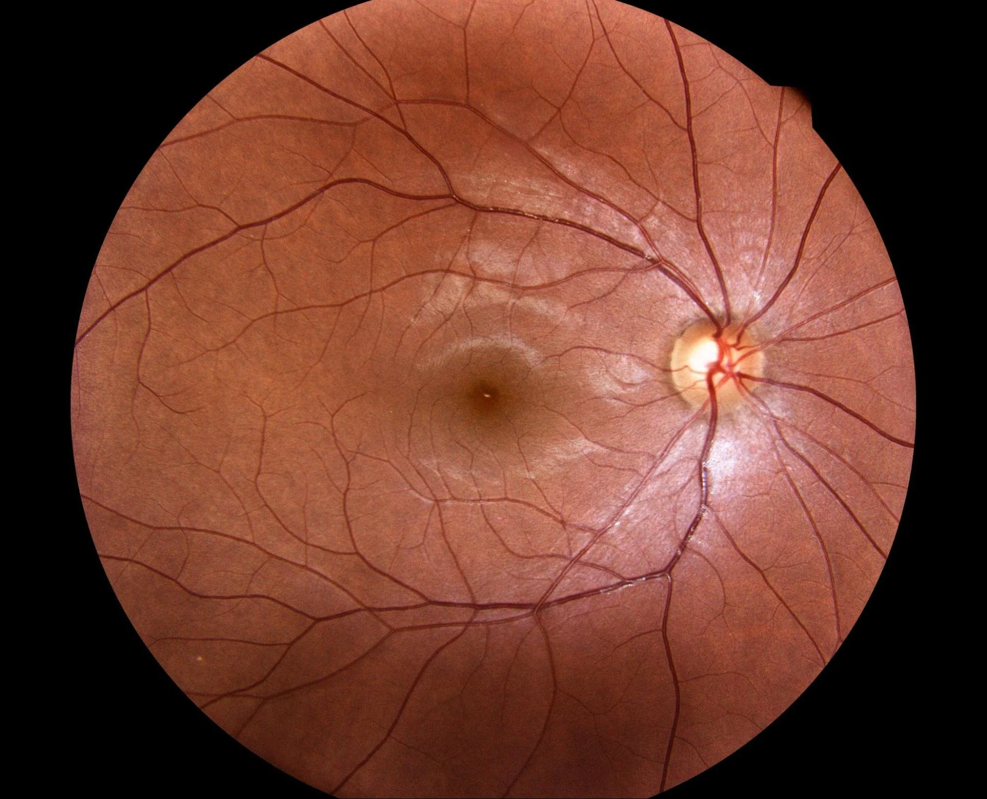
The retina as seen through an ophthalmoscope. The round structure to the right of the photo is the optic disc. The dark structure in the center of the photo is the macula.
When light enters your eye, it comes to a focus on your retina. The retina is a highly specialized group of cells that sends an electrochemical signal to your brain for further processing.
Among all of the cells in the retina, the rods and cones are certainly two of the most important. They absorb vitamin A from all the carrots and green leafy vegetables that you eat. The vitamin A combines with a protein called opsin. The combination is then called rhodopsin or visual purple. A photon of light slams into the rhodopsin molecule thereby sending the neural signal to the brain.
When you look at an object, you are seeing it with the central portion of your retina called the macula. In the center of the macula there is a very small area which allows your brain to discern tiny objects; this is called the fovea. With age, the macula gets less oxygen. If this process continues, and the macula tissue starts to break down, then the disease is called macular degeneration.
Optic Nerve
You might be wondering how visual information gets to the brain from the retina. Like most nerve cells, the retinal ganglion cells have structures called axons. These are ultra-thin cables that send light information to the brain. The group of axons that perform this task is called the optic nerve.
During an eye exam, eye doctors love to look inside your eye.
What are they looking at?
Lots of things. Most importantly, we observe where the optic nerve enters your eye. This is called the optic disc. By looking at the optic disc, we can assess your eye health and determine if you have diseases such as glaucoma.
Primary Visual Cortex
If, like most people, you think you see with your eyes, then you would be wrong.
Light energy is gathered by the eyes and then transformed into an electrochemical signal that is sent to the brain for visual processing – otherwise known as “sight”. So we see with our “brains”, not with our “eyes”.
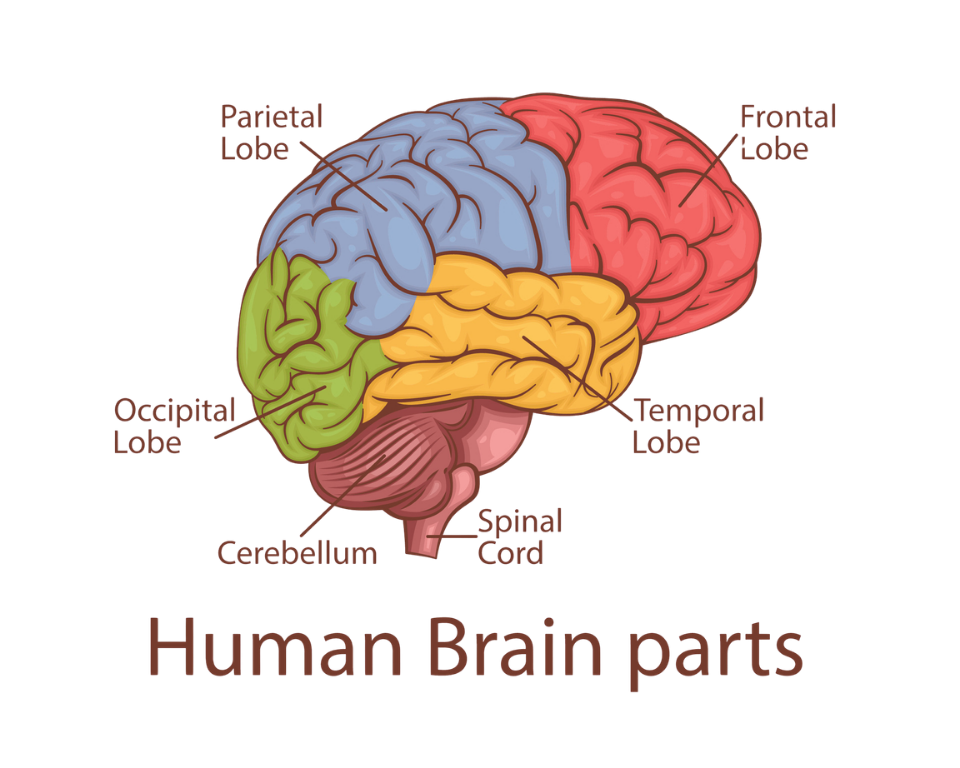
Nerve fibers from the retina are sent to the brain via the visual pathway. The final destination for these fibers? The primary visual cortex (PVC)which is located in the occipital lobe of the brain (see the illustration above). The network of nerve cells and fibers in the PVC create a visual image from the retinal neural data.
Nerve cells in the PVC are activated by visual stimuli such as movement, contrast, depth, and orientation of objects. At the same time the PVC communicates with surrounding visual areas that are specialized for detecting specific features of our visual world. The PVC then gathers information from other areas of the brain for higher level processing of information. This complex neural input provides information for specific object recognition while utilizing our previous visual experience.
Complicated? You betcha! In future blogs, I will present topics in more detail and give real-world examples to help you understand the complexity and beauty of vision.